Discover how a Novel Photoacoustic Contrast Agent for Tumour in humans offers a cost-effective, non-invasive alternative to PET scans, revolutionizing cancer diagnostics.
Introduction
Cancer remains one of the world’s most formidable health challenges, with early and accurate detection being crucial for effective treatment. Traditional imaging techniques like Positron Emission Tomography (PET) have long been the gold standard for visualizing tumours, but they come with significant limitations, including high costs and exposure to radiation. In a groundbreaking development, researchers at the Indian Institute of Science (IISc) have introduced a novel photoacoustic contrast agent for tumour imaging in humans. This innovation promises to transform cancer diagnostics by offering a minimally invasive, cost-effective, and highly sensitive alternative to existing methods.
This article delves deep into the science, significance, and future potential of this new photoacoustic contrast agent, integrating expert insights, recent statistics, and practical information for patients, clinicians, and researchers alike.
The Science Behind Photoacoustic Imaging
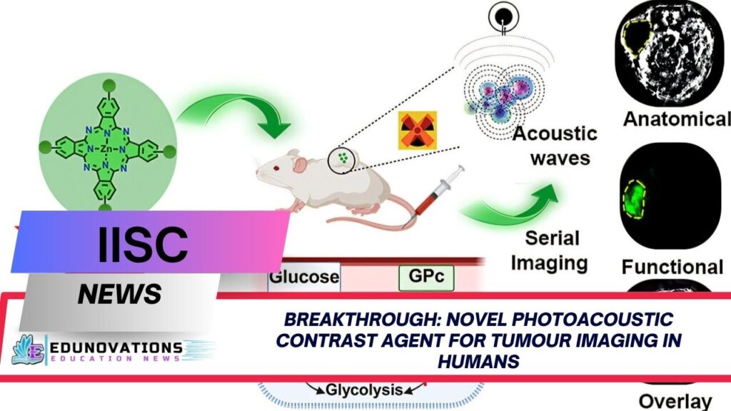
Photoacoustic imaging is an emerging technology that combines the advantages of optical and ultrasound imaging. It involves the use of a near-infrared (NIR) laser beam to excite light-absorbing molecules, known as chromophores, which are introduced into the body. When these molecules absorb the laser energy, they expand and generate pressure waves, which are then detected as sound signals. These signals are analyzed to create detailed three-dimensional images of tissues, allowing clinicians to visualize tumours with remarkable clarity.
How Does the Novel Photoacoustic Contrast Agent Work?
The novel photoacoustic contrast agent for tumour imaging in humans developed at IISc is a biocompatible small molecule designed specifically for this purpose. The agent, referred to as GPc, consists of four glucose units conjugated to a zinc-phthalocyanine scaffold. This unique structure enables the agent to target tumour cells, which have a higher metabolic activity and consume more glucose than healthy cells.
- Targeted Uptake: Tumour cells naturally uptake more glucose, allowing the contrast agent to accumulate preferentially at tumour sites.
- Enhanced Solubility: The glucose units increase the solubility of the molecule, ensuring efficient delivery to the target tissue.
- Non-Invasive Detection: The agent enables non-invasive, high-resolution imaging of superficial tumours, reducing the need for surgical biopsies.
Toppers Use Mind Maps to score more than 95%
-
NCERT Class 11th Commerce Mind Maps
Add to cartOriginal price was: ₹999.00.₹199.00Current price is: ₹199.00. -
NCERT Class 12th Chemistry Mind Maps
Add to cartOriginal price was: ₹199.00.₹75.00Current price is: ₹75.00. -
NCERT Class 12th Commerce Mind Maps
Add to cartOriginal price was: ₹999.00.₹199.00Current price is: ₹199.00. -
NCERT Class 12th Science Mind Maps
Add to cartOriginal price was: ₹999.00.₹199.00Current price is: ₹199.00. -
NCERT Mind Maps For Class 10th
Add to cartOriginal price was: ₹999.00.₹199.00Current price is: ₹199.00.


Purchase Today
Advantages Over Conventional Imaging Techniques
Cost-Effective Alternatives to PET Scan for Superficial Tumour Detection
One of the most significant advantages of this new approach is its potential as a cost-effective alternative to PET scan for superficial tumour detection. PET imaging, while highly effective, is expensive and involves the use of radioactive tracers, which can pose risks with repeated exposure.
Key Benefits:
- Lower Cost: The materials and procedures involved are significantly less expensive than PET or MRI.
- No Radiation Risk: Unlike PET, the photoacoustic method does not expose patients to harmful radiation.
- Minimally Invasive: The procedure requires only a simple injection of the contrast agent, followed by imaging with a NIR laser.
Glucose-Conjugated Zinc-Phthalocyanine: The Science Explained
The core of this innovation lies in the use of glucose-conjugated zinc-phthalocyanine for non-invasive tumour imaging. Zinc-phthalocyanine is a molecule known for its excellent light absorption in the NIR range, making it ideal for photoacoustic imaging. By attaching glucose units, the researchers have created a molecule that is not only highly effective at targeting tumours but also safe and biocompatible.
Mechanism of Action
- Selective Uptake: The glucose units exploit the increased glucose metabolism of tumour cells, ensuring that the agent accumulates primarily in cancerous tissues.
- Stable and Non-Toxic: The agent is designed to be stable in the body and does not interfere with normal cellular processes.
- Clear Imaging: The strong NIR absorption of zinc-phthalocyanine provides clear, high-contrast images of tumour sites.
Minimally Invasive Photoacoustic Tomography for Cancer Diagnosis
The application of minimally invasive photoacoustic tomography for cancer diagnosis represents a significant leap forward in the field of medical imaging. This technique allows for the real-time visualization of tumours without the need for invasive procedures or exposure to radiation.
Clinical Implications
- Early Detection: Enables the identification of tumours at an early stage, improving the chances of successful treatment.
- Monitoring Treatment: Allows clinicians to monitor the effectiveness of therapies in real-time.
- Patient Comfort: Reduces discomfort and anxiety associated with traditional imaging methods.
New Biocompatible Small Molecule Agents for 3D Tumour Visualization
The development of new biocompatible small molecule agents for 3D tumour visualization is a testament to the rapid advancements in bioengineering and medical research. These agents are designed to be safe, effective, and easy to use, making them suitable for widespread clinical adoption.
Features of the New Agent
- High Sensitivity: Capable of detecting even small tumours with high accuracy.
- 3D Imaging: Provides detailed three-dimensional images, aiding in precise diagnosis and treatment planning.
- Biocompatibility: Minimizes the risk of adverse reactions or toxicity.
Expert Insights and Industry Perspectives
In a statement, Dr. Sanhita Sinharay, Assistant Professor at the Department of Bioengineering, IISc, and corresponding author of the study, highlighted the significance of this breakthrough:
“You are able to use a more cost-effective technique, cheaper than both PET and Magnetic Resonance Imaging (MRI), and get the same information. This could fundamentally change how we diagnose and monitor tumours, especially in settings where resources are limited.”
Dr. Pooja Patkulkar, the study’s first author, added:
“One of the major milestones for us was the mechanistic evaluation of the probe. We wanted to see whether the molecule we made was being taken up by the glucose transporters, and what its fate was after uptake. The results were promising and pave the way for further clinical studies.”
The Road Ahead: Future Applications and Research
While the initial results are promising, further research is needed to fully understand the long-term safety and efficacy of this novel photoacoustic contrast agent for tumour imaging in humans. Ongoing studies aim to evaluate its performance in larger clinical trials and explore its potential for detecting other types of cancer.
Potential Future Directions
- Expansion to Other Cancers: Investigating the agent’s effectiveness in imaging other cancer types.
- Integration with AI: Using artificial intelligence to enhance image analysis and diagnostic accuracy.
- Global Accessibility: Developing cost-effective production methods to make the technology accessible worldwide.
Statistics: The Need for Innovation in Cancer Imaging
- According to the World Health Organization, cancer is responsible for nearly 10 million deaths annually worldwide.
- Early detection can increase the five-year survival rate for many cancers by up to 90%.
- PET scans, while effective, can cost upwards of $5,000 per procedure, limiting accessibility in low-resource settings.
- The novel photoacoustic contrast agent for tumour imaging in humans has the potential to reduce imaging costs by up to 70%, according to preliminary estimates from the IISc research team.
Practical Information for Patients and Clinicians
What Does This Mean for Patients?
- Safer Diagnostics: Reduced risk of radiation exposure.
- Affordable Care: Lower costs make advanced imaging accessible to more patients.
- Faster Results: Real-time imaging enables quicker diagnosis and treatment decisions.
For Healthcare Providers
- Easy Integration: The technology can be incorporated into existing imaging workflows with minimal training.
- Enhanced Accuracy: Improved sensitivity and specificity aid in better treatment planning.
Related Resources
- NCERT Courses
- Current Affairs
- Notes
- MCQ’s
- Videos
- Syllabus
- Free NCERT PDFs
- NCERT Mind Maps
- Need Website for Schools? Contact Mart India Infotech
Frequently Asked Questions (FAQs)
1. What is a novel photoacoustic contrast agent for tumour imaging in humans?
A novel photoacoustic contrast agent for tumour imaging in humans is a specially designed molecule that enhances the visibility of tumours during photoacoustic imaging, enabling non-invasive and accurate cancer detection.
2. How does the new agent compare to PET scans?
It offers a cost-effective alternative to PET scan for superficial tumour detection, with no radiation exposure and lower costs.
3. What is glucose-conjugated zinc-phthalocyanine used for in cancer diagnosis?
Glucose-conjugated zinc-phthalocyanine is used for non-invasive tumour imaging, leveraging the high glucose uptake of cancer cells to provide targeted imaging.
4. Is photoacoustic tomography minimally invasive?
Yes, minimally invasive photoacoustic tomography for cancer diagnosis requires only a simple injection of the contrast agent and uses NIR laser imaging.
5. Are there any risks associated with the new biocompatible small molecule agents?
Current studies indicate these agents are biocompatible and safe, but further research is ongoing to confirm long-term safety.
6. Can this technology be used for all types of tumours?
While particularly effective for superficial tumours, ongoing research is exploring its use for other cancer types.
7. How accurate is 3D tumour visualization with this agent?
The new biocompatible small molecule agents for 3D tumour visualization provide high-resolution, detailed images for precise diagnosis.
8. What are the benefits for patients?
Patients benefit from safer, more affordable, and faster diagnostic procedures compared to traditional methods.
9. How soon will this technology be available in clinics?
Clinical adoption depends on the results of ongoing trials and regulatory approvals, but initial results are promising.
10. Where can I find more resources on cancer imaging and diagnostics?
You can explore NCERT Courses, Current Affairs, Notes, and more for in-depth learning.