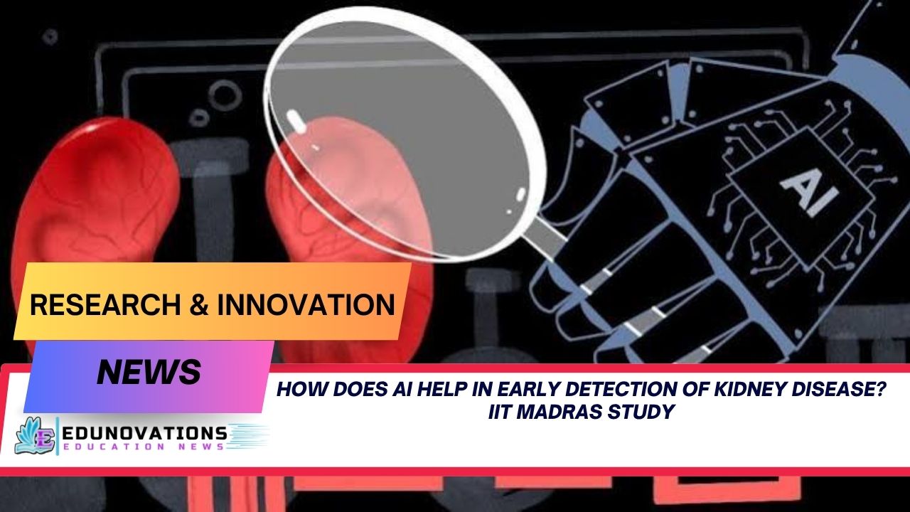
Discover how does ai help in early detection of kidney disease through breakthrough tools created by IIT Madras and CMC Vellore researchers in medical imaging.
Chronic kidney disease (CKD) represents one of the most silent yet devastating health crises facing global healthcare infrastructure today. Because early-stage renal degradation frequently manifests without overt clinical symptoms, millions of individuals remain entirely unaware of their deteriorating organ function until irreversible physiological damage occurs. Addressing this silent epidemic demands diagnostic modalities that move beyond traditional reactive testing toward predictive precision. In a major technological milestone, a collaborative team of researchers from the Indian Institute of Technology Madras (IIT Madras) and Christian Medical College (CMC), Vellore, has unveiled a suite of artificial intelligence framework platforms engineered to transform renal care.
To understand the broader implications of these clinical innovations, healthcare practitioners must examine a fundamental question: how does ai help in early detection of kidney disease across diverse healthcare environments? By unifying routine laboratory variables, automated radiographic image triage, and structural three-dimensional volume quantification, researchers are establishing a new paradigm for rapid diagnostic workflows.
The Growing Crisis of Silent Renal Degradation
Renal pathology progresses through subtle biochemical and architectural alterations that often evade timely clinical capture. In typical clinical settings, patients are evaluated only after presenting systemic complaints such as fluid retention, severe hypertension, or marked fatigue. By this stage, glomerular filtration rates have often fallen dramatically, leaving limited therapeutic avenues beyond intensive pharmaceutical management, ongoing dialysis, or organ transplantation.
The financial and operational strain on national health systems is severe. Dialysis regimens impose massive recurring expenses on families and public medical insurance frameworks. Furthermore, primary care clinics in underserved or rural regions frequently lack specialized nephrologists who can perform granular evaluations of complex abdominal computed tomography (CT) scans.
Integrating intelligent clinical algorithms directly into the diagnostic pipeline effectively bridges this expertise gap. Automated diagnostic assistance acts as a continuous digital screen, flagging high-risk individual profiles long before acute symptoms compel emergency hospital admissions.
Decoding the Breakthrough: Three Interconnected AI Technologies
The collaborative innovation spearheaded by Prof. G.L. Samuel from the Department of Mechanical Engineering at IIT Madras, research scholar Ms. Jennifer Delighta (IIT Madras), and Prof. Santosh Varughese from the Department of Nephrology at CMC Vellore introduces a multi-tiered technological framework. Rather than relying on a single isolated algorithm, the researchers constructed three distinct systems that operate synergistically to evaluate renal health from preliminary screening to surgical mapping.
       +-------------------------------------------------------+
       |           Patient Data & Medical Imaging              |
       +-------------------------------------------------------+
                                   |
         +-------------------------+-------------------------+
         |                         |                         |
         v                         v                         v
+------------------+     +-------------------+     +-------------------+
|  Clinical Data   |     |   2D CT Scans     |     |   3D Imaging      |
|  ML Model        |     |   Deep Learning   |     |   Framework       |
+------------------+     +-------------------+     +-------------------+
         |                         |                         |
         v                         v                         v
+------------------+     +-------------------+     +-------------------+
|  Predicts CKD    |     |  Classifies Cysts,|     |  Measures Volume  |
|  Risk Score      |     |  Stones & Tumors  |     |  & Organ Burden   |
+------------------+     +-------------------+     +-------------------+
         |                         |                         |
         +-------------------------+-------------------------+
                                   |
                                   v
       +-------------------------------------------------------+
       |             Holistic Kidney Digital Twin              |
       +-------------------------------------------------------+
1. Clinical Risk Prediction Engine
The initial tier consists of a machine learning architecture built to evaluate routine clinical and laboratory datasets. Trained using multi-variable records containing 26 discrete biochemical parameters—including serum creatinine, blood urea nitrogen, urine specific gravity, glucose metrics, and blood pressure indicators—the model computes a precise individual risk profile for chronic kidney disease.
By applying advanced ensemble techniques, including Random Forest classifications, the platform calculates hidden probability scores for early renal compromise. This enables general practitioners working in basic medical centers to run routine blood panel outputs through a prototype interface and instantly determine whether a patient requires specialized nephrological follow-up.
2. Automated Deep Learning CT Classifier
The second technology transitions from numerical biomarker analysis to direct radiographic triage. The research team constructed a deep convolutional neural network trained on a vast repository of over 12,000 abdominal CT scans. This vision model reads, segments, and categorizes cross-sectional kidney scans within seconds.
The deep learning system classifies input scans into four specific operational diagnostic categories:
- Normal Kidney: Verifies healthy tissue architecture without structural anomalies.
- Kidney Cyst: Flags fluid-filled structural sacs requiring routine monitoring.
- Kidney Stone: Detects nephrolithiasis density formations, supporting timely intervention.
- Kidney Tumor: Identifies suspicious solid lesions demanding urgent oncological assessment.
3. Patient-Specific 3D Anatomical Reconstruction
The third component is an open-source, three-dimensional imaging framework that converts standard two-dimensional CT slice stacks into volumetric organ models. Standard radiologic reports typically estimate lesion magnitude through linear planar measurements (length times width), which often understate or overestimate complex, irregularly shaped masses.
By generating a complete 3D volumetric rendering of the patient’s individual kidney, this platform calculates exact total organ volume, isolate precise tumor burden, and determines the percentage of surrounding parenchymal tissue affected. Such refined visual metrics provide surgeons with critical spatial awareness before performing partial or radical nephrectomies.
Technological Comparison: AI Diagnostics vs. Traditional Workflows
To understand the practical impact of these computational platforms, consider how automated multi-modal analysis compares against conventional manual evaluation across key diagnostic metrics:
| Metric / Parameter | Traditional Diagnostic Workflow | IIT Madras & CMC Vellore AI Framework |
| Primary Data Input | Sequential manual lab reviews & individual CT reads | Integrated clinical lab metrics + automated CT analysis |
| Early CKD Triage | Dependent on clinical presentation & manual score calculation | Automated machine learning risk evaluation across 26 parameters |
| CT Analysis Speed | 15–45 minutes per scan series (subject to radiologist queue) | Automated classification into four categories within seconds |
| Tumor Assessment | 2D linear planar approximations (Length × Width) | Volumetric 3D reconstruction measuring exact lesion burden |
| Software Accessibility | Proprietary, high-cost workstation radiology suites | Open-source, flexible, cost-effective computational framework |
| Diagnostic Consistency | Varies based on clinician experience and fatigue levels | Standardized algorithmic output across primary and tertiary centers |
Expert Insights and Clinical Perspectives
The development team emphasized that these digital tools are designed to augment clinician expertise rather than replace human medical decision-making. Commenting on the driving objective behind the project, Prof. G.L. Samuel from the Department of Mechanical Engineering at IIT Madras stated:
“The team aimed to develop intelligent systems that would help clinicians make quicker and more informed decisions. We used machine learning along with clinical knowledge to develop tools that would assist in the earlier detection of kidney diseases and give more detailed information specific to the patient.”
Focusing on the critical clinical window for preventive intervention, research scholar Ms. Jennifer Delighta highlighted the strategic advantage provided by volumetric visual analysis:
“Early detection is of paramount importance when dealing with kidney diseases; these AI tools can help detect at-risk patients early and plan their treatment more effectively. The patient-specific imaging framework is of significant promise as it goes beyond the standard measurements to give a more comprehensive picture of the extent of the disease.”
Leading medical experts in nephrology have similarly noted that combining predictive numerical algorithms with diagnostic computer vision resolves long-standing bottlenecks in rural medical access. By giving general medicine physicians access to standardized screening assistance, regional hospitals can prioritize urgent cases, avoiding diagnostic delays that lead to end-stage renal disease (ESRD).
To explore fundamental biological concepts and core science foundations related to human anatomy and organ systems, students and educators can reference NCERT Courses and review structured study materials via NCERT Mind Maps. Understanding foundational human physiology provides essential context for appreciating how technological innovations are reshaping medical science.
Architectural Deep Dive: Building the “Kidney Digital Twin”
Beyond immediate triage applications, the research supported by institutional funding from IIT Madras and the Scheme for Promotion of Academic and Research Collaboration (SPARC) lays the groundwork for a comprehensive Kidney Digital Twin.
A Digital Twin in modern medicine refers to an adaptive, highly detailed virtual representation of a physical human organ. By continuously updating a digital model with real-time patient lab work, imaging updates, and biometric telemetry, clinicians can simulate disease progression within a virtual environment.
+-----------------------------------------------------------------+
|                       PATIENT REALITY                           |
|  +---------------------+           +-------------------------+  |
|  | Laboratory Panels   |           | Serial Radiographies    |  |
|  +---------------------+           +-------------------------+  |
+-----------------------------------------------------------------+
                               |
                               v (Continuous Data Ingestion)
+-----------------------------------------------------------------+
|                   KIDNEY DIGITAL TWIN SYSTEM                    |
|  +-----------------------------------------------------------+  |
|  |  Volumetric 3D Anatomical Render                          |  |
|  |  + Baseline Clinical Risk Trajectory                      |  |
|  +-----------------------------------------------------------+  |
|                              |                                  |
|                              v                                  |
|  +-----------------------------------------------------------+  |
|  |  In-Silico Simulation Engine                              |  |
|  |  - Test Pharmacological Responses                         |  |
|  |  - Project Glomerular Filtration Rates                    |  |
|  |  - Simulate Surgical Resection Outcomes                   |  |
|  +-----------------------------------------------------------+  |
+-----------------------------------------------------------------+
                               |
                               v
+-----------------------------------------------------------------+
|                   PERSONALIZED CLINICAL ACTION                  |
|  - Optimized Prescriptions     - Minimal Tissue Loss Surgery   |
+-----------------------------------------------------------------+
Key Capabilities of Digital Twin Technology in Nephrology:
- Longitudinal Disease Forecasting: Predictive simulation models plot estimated loss of kidney function over a multi-year horizon, allowing doctors to adjust therapeutic strategies proactively.
- Virtual Therapeutic Testing: Clinicians can evaluate how specific drug dosages or antihypertensive regimens might impact an individual patient’s unique kidney structure before administering treatment.
- Surgical Simulation: Before performing complex tumor resections, surgeons can rehearse cuts on the virtual 3D organ model to maximize healthy tissue preservation.
- Integration with Wearable Sensors: Future phases aim to connect this virtual model with non-invasive wearable sensors that track continuous electrolyte variations, hydration metrics, and metabolic markers in real time.
Medical scholars preparing for competitive academic examinations or staying informed on scientific breakthroughs can access comprehensive preparatory frameworks through the Syllabus Section and review curated subject breakdowns in the Notes Directory.
Step-by-Step Diagnostic Pipeline Implementation
Understanding how these distinct AI layers operate within an integrated hospital software environment highlights the shift toward streamlined clinical workflows:
- Patient Data Ingestion:Routine patient intake gathers demographic information, blood profiles, and urinalysis values. These raw inputs populate the machine learning risk assessment dashboard.
- Algorithmic Risk Scoring:The classification engine computes an initial risk index for chronic kidney disease. If the calculated probability exceeds pre-established safety thresholds, the system flags the profile for radiological imaging.
- Radiographic Scanning & Automated Segmentation:The patient undergoes an abdominal CT scan. The deep learning vision network instantly processes the image files, identifying the presence of structural anomalies such as cysts, renal calculi, or solid masses.
- Volumetric 3D Generation:For scans showing structural lesions or neoplastic growths, the system builds a 3D volumetric organ rendering to calculate precise tumor density and parenchymal tissue distribution.
- Multidisciplinary Decision Support:The attending physician, radiologist, and nephrologist review the consolidated single-page digital report, arriving at a consensus treatment plan in minutes rather than days.
Healthcare institutions and educational organizations looking to deploy robust web infrastructure to present scientific research or streamline administrative workflows can partner with specialized digital development providers like Mart Ind Infotech for customized technical solutions.
Broader Implications for Global Healthcare Systems
The broader deployment of artificial intelligence in public health infrastructure addresses major operational challenges facing healthcare delivery worldwide:
+----------------------------------------------------------------+
|                 GLOBAL HEALTHCARE SYSTEM BENEFITS              |
+----------------------------------------------------------------+
                               |
         +---------------------+---------------------+
         |                                           |
         v                                           v
+---------------------------------+  +---------------------------------+
|   Resource-Constrained Clinics  |  |    Surgical & Tertiary Hubs    |
| - Automated Triage              |  | - Volumetric Precision          |
| - Early Specialist Referral     |  | - Reduced Operative Margin Risk |
| - Decreased Dialysis Dependence |  | - Tailored Oncological Care     |
+---------------------------------+  +---------------------------------+
In developing nations, primary health centers are often located far from major tertiary hospitals. Implementing deep learning models for CT scan kidney stone detection enables remote clinics to screen incoming patients accurately using lower-cost local imaging infrastructure.
When local general practitioners can accurately differentiate benign fluid cysts from aggressive solid tumors, unnecessary and costly emergency transfers to urban centers are minimized. Conversely, high-risk cases receive immediate, targeted specialist referrals, accelerating life-saving interventions.
To keep track of contemporary scientific advancements, educational updates, and technological policy developments, readers can follow real-time analysis through Current Affairs, reinforce key concepts using target practice exercises in MCQ Practice Sets, or watch expert video tutorials via Educational Videos.
Future Roadmap: Multi-Center Validation and Deployment
While the performance metrics achieved by the IIT Madras and CMC Vellore research group are promising, transitioning AI models from academic prototypes to certified clinical medical devices requires rigorous testing.
The research team is expanding its validation roadmap across several key phases:
- Multi-Center Clinical Datasets: Validating algorithm accuracy across diverse demographic cohorts, varied imaging hardware brands, and distinct hospital diagnostic environments.
- Open-Source Tool Standardization: Refining the underlying open-source 3D visualization frameworks so they integrate seamlessly with existing hospital Picture Archiving and Communication Systems (PACS).
- Regulatory Compliance Frameworks: Aligning software performance standards with medical device regulations to ensure absolute patient safety and data confidentiality.
- Wearable Health Monitoring: Exploring long-term integration with non-invasive biochemical sensors to feed continuous metabolic metrics into individual organ digital twin profiles.
As these validation milestones are completed, automated diagnostic platforms will play an increasingly vital role in helping clinicians protect renal health worldwide. Students and educators seeking offline learning resources can access free study content through Free NCERT Downloads.
Toppers Use Mind Maps to score more than 95%
NCERT Class 11th Commerce Mind Maps
Add to cartOriginal price was: ₹999.00.₹199.00Current price is: ₹199.00.NCERT Class 12th Chemistry Mind Maps
Add to cartOriginal price was: ₹199.00.₹75.00Current price is: ₹75.00.NCERT Class 12th Commerce Mind Maps
Add to cartOriginal price was: ₹999.00.₹199.00Current price is: ₹199.00.NCERT Class 12th Science Mind Maps
Add to cartOriginal price was: ₹999.00.₹199.00Current price is: ₹199.00.NCERT Mind Maps For Class 10th
Add to cartOriginal price was: ₹999.00.₹199.00Current price is: ₹199.00.





Purchase Today
Frequently Asked Questions (FAQs)
1. How does AI help in early detection of kidney disease?
AI assists by analyzing subtle biochemical markers in routine lab work and identifying early structural changes in CT scans long before physical symptoms appear. This automated analysis provides clinicians with rapid, standardized risk scores to guide timely medical interventions.
2. What types of data does the machine learning risk model evaluate?
The machine learning model processes standard laboratory and clinical parameters, including serum creatinine levels, blood urea nitrogen, blood pressure metrics, glucose readings, and urine specific gravity to calculate an individual’s chronic kidney disease risk.
3. How does deep learning models for CT scan kidney stone detection work?
The deep learning classifier uses convolutional neural networks trained on over 12,000 CT scans to analyze cross-sectional images automatically. It quickly distinguishes between normal kidney tissue, cysts, stones, and solid tumors.
4. What is a digital twin for kidney disease diagnosis?
A digital twin is a dynamic, patient-specific virtual 3D model of the organ created from CT scans and clinical metrics. It allows doctors to track disease progression over time, simulate surgical procedures, and personalize treatment plans.
5. Can machine learning predict chronic kidney disease risk before symptoms appear?
Yes. Machine learning algorithms detect complex patterns across multiple clinical variables that human observation might miss, allowing doctors to identify at-risk patients during early, asymptomatic stages of renal decline.
6. What makes ai tools for classifying kidney cyst vs tumor CT scan more effective than traditional methods?
Unlike conventional 2D measurements that only evaluate length and width, AI-driven tools build complete 3D volumetric reconstructions. This approach measures exact tumor volume and parenchymal organ involvement with much higher spatial accuracy.
7. Was this AI diagnostic system developed using open-source software?
Yes, the 3D visual reconstruction framework was engineered using flexible, open-source software platforms. This design choice ensures the technology remains accessible and cost-effective for hospitals and research institutions.
8. Is this AI system intended to replace radiologists and nephrologists?
No. The platform is designed as an intelligent clinical decision-support tool. It assists doctors by standardizing initial image triage, shortening evaluation times, and providing detailed volumetric data to guide final clinical decisions.
9. What are the next steps before this technology is deployed in hospitals?
The research team is expanding clinical validation across larger, multi-center patient datasets, ensuring software compatibility with standard hospital PACS platforms, and meeting regulatory medical device standards.
10. How might wearable technology integrate with kidney AI tools in the future?
Future iterations aim to link non-invasive wearable sensors directly into the patient’s digital twin model. This connection will provide continuous monitoring of key metabolic markers and fluid balance outside hospital settings.